Glossary of Vision Terms
Excerpted in part from “Dictionary of Eye
Terminology” © 1990-2003 by Triad Communications
|
Click HERE for eye diagram |
|
|
|
|
|
ACCOMMODATION |
The ability of the eye to quickly change its focus from distant to near objects. Process achieved by the internal lens changing its shape. |
|
ALLERGIES |
Reaction which the body undergoes to protect itself from infection overreacts to non-infectious substances. Click HERE for more info. |
|
AMBLYOPIA (lazy eye) |
Poor vision in an eye due to lack of visual attention. May be cause by crossed eye or large difference in clarity or size of image between eyes. Click HERE for more info. |
|
AMSLER GRID |
A special grid (looks like graph paper) in which an individual may determine whether there is any distortion to vision. Given to those who are at risk of macular swelling or neovascularization. |
|
ANGLE |
The area of the eye where the iris and cornea meet. This area contains the trabecular meshwork and is where fluid drains out of the eye. |
|
ANTERIOR CHAMBER |
The space in front of the iris and behind the cornea. Filled with aqueous fluid. Click HERE for eye diagram |
|
AQUEOUS |
Clear, watery fluid that flows within the eye and nourishes the lens and the cornea; secreted by the ciliary processes. |
|
ARCUS |
White ring which develops near the outer edge of the cornea. Com-posed of cholesterol. Those with arcus should have a cholesterol screening to rule out elevated levels, though often arcus may be present even with normal cholesterol. |
|
ASTEROID HYALOSIS |
Small calcium particles that form in the vitreous and may interfere with vision. More common in the elderly. |
|
ASTIGMATISM |
A condition in which the surface of the cornea is not spherical or 'round'; causes a blurred image to be received at the retina. |
|
BAND KERATOPATHY |
Areas of calcium buildup that develop on the cornea. May be a sign of uveitis or gout. |
|
BELL'S PALSY |
Numbness developing on one side of the face and lasting a couple weeks. That side of the face commonly droops. May be caused by a viral infection. |
|
BINOCULAR VISION |
The blending of the separate images seen by each eye into a single image. Allows images to be seen with depth. |
|
BLEPHARITIS |
Inflammation of the eyelids which may cause redness, irritation, and a crustiness along the lid edges. Tends to be a chronic condition. Warm compresses and antibiotic treatment may help. |
|
BLIND SPOT |
(1) A small area of
the retina where the optic nerve enters the eye; occurs normally in all eyes.
There is no vision in this part of the retina. |
|
BULL'S EYE MACULOPATHY |
Term for the appearance of the macula when toxic buildup of certain medications occurs. Chloroquine is one of the more common medications to cause this. |
|
CATARACT |
A cloudiness that develops within the lens of the eye. Click HERE for more info. Click HERE for eye diagram |
|
CELLOPHANE MACULOPATHY |
Also known as epiretinal or preretinal membrane. A film of cells that develops over the retina, which in some cases can interfere with vision permanently. |
|
CELLULITIS |
Swelling and tenderness of an eyelid. Usually caused by irritation or infection. If pain or double vision occur, immediate visit to the eye doctor is necessary to rule out more serious infections. |
|
CENTRAL SEROUS CHORIORETINOPATHY |
Condition that often affects males in their 30s and 40s who are under a lot of stress. Fluid builds up in the macula, resulting in blurred, distorted and shrunken images in that eye compared to the other. May last for several weeks or months. |
|
CENTRAL RETINAL ARTERY |
The blood vessel that carries blood into eye; supplies nutrition to the retina. |
|
CENTRAL RETINAL VEIN |
The blood vessel that carries blood from the retina. |
|
CHALAZION |
Commonly called a stye. Blocked oil gland on edge of eyelid which produces a visible bump. Warm compresses may help unclog the gland. |
|
CHOROID |
The layer of the eye filled with blood vessels that lays behind and nourishes the retina; part of the uvea. Click HERE for eye diagram |
|
CILIARY MUSCLES |
The muscles that pull on the zonules to enable the lens to change shape for focusing. Click HERE for eye diagram |
|
CILIARY PROCESSES |
The extensions or projections of the ciliary body that secrete aqueous humor. |
|
COLOR BLINDNESS |
More accurately called color vision deficiency. Hereditary condition present from birth in 8% of males and .5% of females. Colors are seen differently by these people, and shades of green and red may appear identical. Any change in the appearance of colors noticed later in life should be investigated. |
|
CONES, CONE CELLS |
One type of specialized light-sensitive cells (photoreceptors) in the retina that provide sharp central vision and color vision. Also see RODS. |
|
CONJUNCTIVA |
The thin, moist skin (membrane) that lines the inner surfaces of the eyelids and the outer surface of the sclera. Click HERE for eye diagram |
|
CONJUNCTIVITIS |
Inflammation of the conjunctiva. Click HERE for more info. |
|
CONTRAST SENSITIVITY |
The ability to perceive differences between an object and its background. |
|
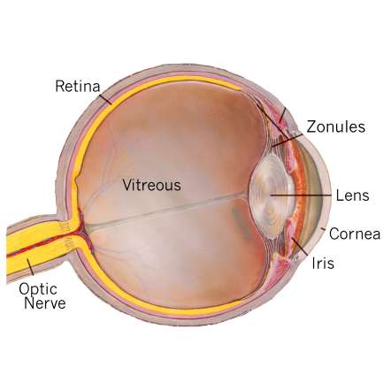
CORNEA |
The outer, transparent, dome-like structure that covers the iris, pupil, and anterior chamber. Part of eye's focusing system. Click HERE for eye diagram |
|
CORNEAL ABRASION |
A scratch of the cornea. Antibiotic treatment will prevent infection. |
|
CORNEAL EROSION |
A sloughing off of the front layer of skin (epithelium) of the eye. This can be caused by injury or hereditary conditions, and usually leaves the eye feeling 'scratchy' or painful. The epithelium re-generates over the course of a couple days and the symptoms go away. |
|
CORNEAL ULCER |
An 'eating away' of the cornea, usually by bacteria. Click HERE for more info. |
|
COTTON-WOOL SPOTS |
Cloudy areas that develop in the retina from a lack of oxygen. May be due to diabetes, carotid artery disease, or other conditions. |
|
CRYSTALLINE LENS |
See 'Lens'. |
|
DEPOSITS |
Debris that builds up on aging contact lenses and interferes with vision and comfort. Usually protein or fat, but may also be makeup and hairspray. |
|
DIABETIC RETINOPATHY |
A condition occurring from Diabetes where the retina shows signs of damage. Click HERE for more info. |
|
DILATION |
A process by which the pupil is temporarily enlarged with special eye drops (mydriatic); allows the eye doctor to better view the inside of the eye. |
|
DOUBLE VISION (diplopia) |
Seeing 'double' is usually due to an over or underaction of one of the muscles that surround an eye. This does not allow both eyes to point in the same direction. Any sudden appearance of double vision should be investigated by an eye doctor. |
|
DRUSEN |
Tiny yellow or white deposits in the retina or optic nerve head. Can potentially be an early sign of Macular Degeneration. |
|
DRY EYE |
A common problem. May be due to dry air, poor tear quality or quantity, blinking infrequently, or a disorder of the eye's surface. Click HERE for more info. |
|
DUANE'S SYNDROME |
A condition where eyes cannot properly turn inward or outward. Often, the eyelids will partially close (because the eye retracts further into the socket) when eye movements are made. |
|
ECTROPION |
Condition, usually in the elderly, in which the eyelids don't stay positioned against the eye. May lead to sever dry eye and uncontrolled tearing. |
|
ENTROPION |
Opposite of ectropion. Eyelids rotate back toward eye, which may cause the eyelashes to scratch the cornea. |
|
EPIRETINAL MEMBRANE |
Also known as preretinal mem-brane or cellophane maculopathy. A film of cells that develops over the retina, which in some cases can interfere with vision permanently. |
|
EPISCLERITIS |
Inflammation of the white of the eye. Blood vessels become very distinct and noticeable. |
|
EPITHELIAL BASEMENT MEMBRANE DYSTROPHY |
EBMD for short. A common condition of the cornea caused by injury or heredity, which produces occasional symptoms of dryness, scratchiness, and pain. Also called 'map-dot-fingerprint dystrophy' because of its appearance. |
|
EXTRAOCULAR MUSCLES |
The six muscles that attach to each eye and rotate the eye into any of the 'positions of gaze'. Any over or underactivity of a muscle will result in double vision (diplopia). |
|
EXUDATES |
Fat products that seep from blood vessels in the retina. Usually caused by diabetes or high blood pressure. |
|
FARSIGHTEDNESS |
Hyperopia; ability to see distant objects more clearly than close objects; may be corrected with glasses, contact lenses, or LASIK. |
|
FLASHES |
Any flashes in vision should be investigated by an eye doctor. Most are due to a tugging on the retina by the vitreous gel and are relatively harmless. However, a detached retina may also produce flashes and this is an emergency. |
|
FLOATERS |
Gray or black specks that appear to drift across one's vision. Most are harmless debris trapped on the inside of the eye. Any new floaters that have suddenly appeared should be immediately examined to rule out the possibility of hemorrhage or retinal detachment. |
|
FLOPPY EYELID SYNDROME |
Condition affecting mostly overweight individuals in which the eyelids are lax and easily flip inside out during sleep. This causes irritation and redness since the inside of the lid rubs on bedsheets. |
|
FLUORESCEIN ANGIOGRAPHY |
A test to examine blood vessels in the retina, choroid, and iris. A special dye is injected into a vein in the arm and pictures are taken as the dye passes through blood vessels in the eye. |
|
FOVEA |
The central part of the macula that provides the sharpest vision. |
|
FUCHS' DYSTROPHY |
Disorder of the back surface of the cornea. Click HERE for more info. |
|
FUNDUS |
The interior lining of the eyeball, including the retina, optic disc, and macula; portion of the inner eye that can be seen during an eye examination by looking through the pupil. Click HERE for eye diagram |
|
GIANT PAPILLARY CONJUNCTIVITIS (GPC) |
A condition from contact lens wear in which spongy bumps form under the upper lids. Due to allergies from contact lens deposits. Disposable lenses improve this situation. |
|
GLAUCOMA |
Eye disease where sensitive retinal nerve fibers are damaged, leading to loss of vision. The damage can occur from high internal pressure or poor blood flow to the nerve fibers. Click HERE for more info. |
|
GRAVES' DISEASE |
Also called Thyroid Eye Disease. Staring appearance, dryness, and double vision may result from inflammation of the muscles that surround the eye. Click HERE for more info. |
|
GUTTATA |
Dimpling of the rear surface of the cornea, which may interfere with clarity of vision in some cases. Fairly common. More numerous guttata are seen in Fuchs' Dystrophy, a hereditary corneal disorder. |
|
HISTOPLASMOSIS |
Infection by a fungus which may cause retinal damage. Click HERE for more info. |
|
HYPEROPIA |
Farsightedness; ability to see distant objects more clearly than close objects; may be corrected with glasses, contact lenses, or LASIK. |
|
HYPERTENSIVE RETINOPATHY |
The retinal manifestations of severe high blood pressure. Vascular changes, exudates, and hemorrhages. |
|
INFILTRATES |
White blood cells accumulating on the cornea to fight infection. May occur from contact lens overwear, even in the absence of infection. |
|
INTRAOCULAR PRESSURE (IOP) |
Pressure of the fluid inside the eye; normal IOP varies among individuals but averages 16 mm Hg. In general, readings above 21 are considered suspicious for glaucoma. |
|
IRIS |
The colored ring of muscle tissue suspended behind the cornea and immediately in front of the lens; regulates the amount of light entering the eye by adjusting the size of the pupil. Click HERE for eye diagram |
|
IRITIS |
Inflammation of the iris. Click HERE for more info. |
|
ITCHY EYES |
Typically due to allergy. The source of the allergy should be determined to best resolve the itchiness. Dry eyes will intensify the itchiness. |
|
KERATITIS |
Inflammation of the cornea. |
|
KERATOCONUS |
Hereditary condition in which the cornea become misshapen, distorting a person's vision. Click HERE for more info. |
|
KRUKENBERG SPINDLE |
A pattern of pigment granules deposited on the posterior surface of the cornea. See 'pigmentary dispersion'. |
|
LACRIMAL DUCT OBSTRUCTION |
Seen in babies, where the tear drainage duct fails to open by birth. Causes excessive tearing since normal tear cannot drain properly. Often resolves on its own or with massage. May require instrument probing to unblock. |
|
LACRIMAL GLAND |
The small almond-shaped structure that produces tears; located just above the outer corner of the eye. |
|
LASIK |
An acronym for 'Laser ASsisted In-situ Keratomiliusis'. Laser surgery for the correction of refractive errors. Click HERE for more info. |
|
LAZY EYE |
See 'AMBLYOPIA' |
|
LEGAL BLINDNESS |
In the U.S: 1.
visual acuity of 20/200 or worse in the better eye with
corrective lenses (20/200 means that a person must be at 20 feet from an eye
chart to see what a person with normal vision can see at 200 feet) or 2.
visual field restricted to 20 degrees
diameter or less (tunnel vision) in the better eye. NOTE: These criteria are
used to determine eligibility for government disability benefits and do not
necessarily indicate a person's ability to function. |
|
LENS |
The transparent, double convex (outward curve on both sides) structure suspended between the aqueous and vitreous; helps to focus light on the retina. Click HERE for eye diagram |
|
LOW VISION |
Visual loss that cannot be corrected with eyeglasses or contact lenses and interferes with daily living activities. |
|
MACULA |
The small, sensitive area of the central retina; provides central vision for fine work and reading. |
|
MACULAR DEGENERATION |
Hereditary condition where the macula becomes scarred and central vision may be lost. Click HERE for more info. |
|
MACULAR EDEMA |
Swelling of the macula. May occur from cataract surgery, trauma, or diabetes. Vision becomes distorted or hazy. |
|
MACULAR HOLE |
Condition where the vitreous gel tugs at the macula and tears a bit of it away. Results in a permanent gray spot in vision directly where one is looking. Often caused by trauma. |
|
MAP-DOT-FINGERPRINT DYSTROPHY |
Also called Epithelial Basement Membrane Dystrophy or EBMD for short. A common condition of the cornea caused by injury or heredity, which produces occasional symptoms of dryness, scratchiness, and pain. |
|
MEIBOMIANITIS |
Inflammation of the meibomian glands, which line the edge of the eyelids. The oily part of the tears are not released, and dry eyes result. |
|
MYASTHENIA GRAVIS |
Double vision or eyelid droop that worsens toward the end of day. May also have other muscle weakness. Caused by viral infection or permanent neurological condition. |
|
MYOKYMIA |
Eyelid twitch usually due to stress or caffeine. More forceful twitching may be due to a parathyroid gland imbalance and is not myokymia. |
|
MYOPIA |
Nearsightedness; ability to see close objects more clearly than distant objects; may be corrected with glasses, contact lenses, or LASIK. |
|
NEARSIGHTEDNESS |
See 'Myopia'. |
|
NEOVASCULARIZATION |
The formation of new blood vessels. Are caused by a lack of oxygen such as with contact lens wear, or conditions like diabetes. They are not good, since they may interfere with vision or bleed easily, which can lead to scarring inside the eye. |
|
NYSTAGMUS |
Rhythmic oscillations of the eyes back and forth. Often caused by poor or no vision early in life. |
|
OCCIPITAL LOBE |
The part of the brain that receives the visual impulses and processes them. Located at the rear of the brain. |
|
OCULAR ISCHEMIC SYNDROME |
Low oxygenation of the retina, usually due to carotid artery disease. May cause hemorrhages and cotton-wool spots. |
|
OCULAR / OPHTHALMIC MIGRAINE |
Type of migraine where one sees vision changes (aura), sometimes followed by headache or nausea. Click HERE for more info. |
|
OPHTHALMOLOGIST |
An ophthalmologist is a medical doctor or doctor of osteopathy who has attended college and four years of medical school with residency. Ophthalmologists are specialists in eye surgery, though most also perform routine eye exams and some fit contact lenses. |
|
OPTIC CUP |
The white, cup-like area in the center of the optic disc. |
|
OPTIC DISC / OPTIC NERVE HEAD |
The circular area (disc) visible inside the eye where the optic nerve connects to the retina. |
|
OPTIC NERVE |
The bundle of over one million nerve fibers that carry visual messages from the retina to the brain. Click HERE for eye diagram |
|
OPTIC NEURITIS |
Inflammation of the optic nerve, sometimes caused by Multiple Sclerosis or viral infection. Click HERE for more info. |
|
OPTICIAN |
An optician is a person trained in the selection, manufacture, and dispensing of eyeglasses and contact lenses. The American Board of Opticianry tests and certifies opticians. |
|
OPTOMETRIST |
A Doctor of Optometry is an eye doctor who has graduated from college and then graduated from a four -year school of optometry. An optometrist can examine eyes and prescribe eyeglasses, contact lenses, and eye medications. Optometrists remove foreign bodies from the eyes, but generally do not perform surgery. |
|
PERIPHERAL VISION |
Side vision; ability to see objects and movement outside of the direct line of vision. |
|
PHOTOPHOBIA |
Light sensitivity. May be due to any number of causes, including infection, corneal abrasion, migraine, or cataract. |
|
PIGMENTARY DISPERSION |
Condition where pigment granules are sloughed off the iris and end up in the angle or posterior corneal surface. Potentially could lead to glaucoma from clogging of the interior drainage system of the eye. Click HERE for eye diagram |
|
PINGUECULA |
Appears as a small yellow bump on the white of the eye, usually toward the nose. A non-cancerous thickening of the skin. May get irritated at times. See your eye doctor to rule out more serious possibilities. |
|
PINK EYE |
Inflammation of the conjunctiva (conjunctivitis) due to either bacteria, viruses, or allergies. May or may not be contagious. Click HERE for more info. |
|
POSTERIOR CHAMBER |
The space between the back of the iris and the front face of the vitreous; filled with aqueous fluid. Click HERE for eye diagram |
|
PRESBYOPIA |
The gradual loss of the eye's ability to change focus (accommodation) for seeing near objects caused by the lens becoming less elastic; associated with aging; occurs in almost all people over age 40. |
|
PSEUDOTUMOR CEREBRI |
High intracranial pressure which is observed by viewing the pushing forward of the optic nerve head. Patient may have headaches and transient vision changes lasting only seconds at a time. Tends to affect females who have had some recent weight gain. Pregnancy and certain medications may also trigger it. |
|
PTERYGIUM |
A thickening of the skin of the white of the eye that starts to grow onto the cornea. Usually due to many years of exposure to the sun. Can be surgically removed if interfering with vision. |
|
PTOSIS |
An eyelid droop. Any sudden onset should be investigated by an eye doctor. |
|
PUNCTAL OCCLUSION |
Short procedure where the punctum is plugged or cauterized to prevent drainage of tears. Performed in those with very dry eyes. |
|
PUNCTUM |
Small drainage opening at the inside corner of each eyelid. Where tears normally drain away. |
|
PUPIL |
The black part of the eye. This is actually an opening that light passes through on its way to the retina at the back of the eye. Click HERE for eye diagram |
|
RADIAL KERATOTOMY (RK) |
The original type of refractive surgery (prior to laser surgery) that used eight spoke-like incisions to induce flattening of the cornea and correct nearsightedness. |
|
REFRACTION |
A test to determine the best eyeglasses or contact lenses to correct a refractive error. |
|
REFRACTIVE ERROR |
Nearsightedness (myopia), farsightedness (hyperopia), and astigmatism. Click HERE for more info. |
|
RETINA |
The light-sensitive layer of tissue that lines the back of the eyeball; sends visual messages through the optic nerve to the brain. Click HERE for eye diagram |
|
RETINAL ARTERY / VEIN OCCLUSION |
Due to clotting or cholesterol problems. Similar to a heart attack, but occurring in the retinal vessels. Results in loss of part of the peripheral vision or occasion-ally complete loss of vision in an eye. |
|
RETINAL DETACHMENT |
Condition where the retina is torn from the back of the eye. Requires immediate attention to prevent blindness. Click HERE for more info. |
|
RETINAL PIGMENT EPITHELIUM (RPE) |
The pigment cell layer that lays behind the retinal cells; located just outside the retina and attached to the choroid. Improves quality of vision by cutting down on reflections within the eye. |
|
RETINITIS PIGMENTOSA |
Hereditary condition in which a person (usually in teen years) notices worsening night vision and then a loss of peripheral vision. Blindness often is the end result. No cure, though vitamins may slow its progression. |
|
RETINOBLASTOMA |
Highly lethal tumor that tends to develop in the retinas of infants and young children. Can be hereditary and also spontaneous. Early treatment can be a life saver. |
|
RETINOPATHY OF PREMATURITY |
Affects infants born more than 2 months prematurely and who are placed in high oxygen environ-ments. Peripheral retina is abnormal and susceptible to detachment and other problems. |
|
RETINOSCOPY |
Viewing of reflections off the retina to determine the person's prescrip-tion. Helpful for small children or those who can't communicate with the optometrist. |
|
RODS, ROD CELLS |
One type of specialized light-sensitive cells (photoreceptors) in the retina that provide side vision and the ability to see objects in dim light (night vision). Also see CONES. |
|
ROSACEA |
A skin condition similar in appearance to acne, but usually affecting older people. A redness to cheeks and nose may develop, and dry eyes are fairly common with this condition. |
|
SCHLEMM'S CANAL |
The passageway for the aqueous fluid to leave the eye. |
|
SCLERA |
The tough, white, outer layer (coat) of the eyeball; with the cornea, it protects the entire eyeball. Click HERE for eye diagram |
|
SCLERITIS |
Inflammation of the interior layers of the sclera. Typically a painful, recurring condition, and often linked to a type of arthritis. NSAIDs or steroids may help. |
|
STARGARDT'S DISEASE |
Hereditary condition in which central vision is slowly lost. Similar to macular degeneration but occurs in children and teens. No treatment is helpful. |
|
STRABISMUS |
Crossed eyes. |
|
SUPERFICIAL PUNCTATE KERATOPATHY (SPK) |
Small irritated areas on the cornea. Visible with the microscope and usually due to dryness or foreign body. |
|
THYROID EYE DISEASE |
Also called Graves' Disease. Staring appearance, dryness, and double vision may result from inflammation of the muscles that surround the eye. Click HERE for more info. |
|
TONOMETRY |
The standard to determine the fluid pressure inside the eye (intraocular pressure). |
|
TOXOPLASMOSIS |
Retinal lesion associated with overlying vitreous haze. Vision loss may occur depending on the position and extent of the lesion. Contracted from cats. |
|
TRABECULAR MESHWORK |
The spongy, mesh-like tissue near the front of the eye that allows the aqueous fluid (humor) to flow to Schlemm's canal then out of the eye through ocular veins. |
|
TWITCHING LIDS |
Eyelid twitching (Myokymia) is common and is usually due to stress or caffeine. More forceful twitching may be due to a parathyroid gland imbalance and is not the same as myokymia. |
|
UVEA, UVEAL TRACT |
The middle coat of the eyeball, consisting of the choroid in the back of the eye and the ciliary body and iris in the front of the eye. |
|
UVEITIS |
Inflammation of the Uvea. Click HERE for more info. |
|
VISUAL ACUITY |
The ability to distinguish details and shapes of objects; also called central vision. |
|
VISUAL FIELD |
The entire area that can be seen when the eye is forward, including peripheral vision. |
|
VITREOUS |
The transparent, colorless mass of gel that lies behind lens and in front of retina. Click HERE for eye diagram |
|
XANTHELASMA |
Cholesterol deposits usually seen as yellow, raised patches under the skin to the sides of the nose. A sign that cholesterol levels may be high. |
|
ZONULES |
The fibers that hold the lens suspended in position and enable it to change shape during accommodation. Click HERE for eye diagram |
|
|
|